| |
 |
over time in brain volumes between groups, multiple general linear modeling for repeated measures analyses were done for each of the volume measures (i.e. whole brain, hypothalamus, cerebral gray and white matter, and lateral and third ventricles), with group (transsexuals, comparisons), sex (male, MF, female, FM) as between-subject factors, and time (scan 2 minus scan 1) as within-subject factors. Age and intracranial volume at first measurement were added as covariates. In case of a significant finding, years of education and scan interval were also added to the statistical analysis as covariates to assess their possible influence. |
The influence of sex hormone treatment on brain morphology in young adult humans between 16 and 45 years of age (on average 25 years) was measured in eight MF and six FM human subjects (Table 1). Highresolution magnetic resonance brain images were obtained prior to and during cross-sex hormone treatment after a 4-month interval and prior to sex reassignment surgery (Fig. 1). MFs received dual therapy: the anti-androgen cyproterone acetate (Androcur) (2!50 mg/day), having anti-androgenic properties both by its progestational action and competing for the androgen receptor, and a synthetic oral estrogen ethinylestradiol (2!50 mg/day). The estrogen doses used are 2–3 times more biopotent than traditional estrogen replacement in women. FMs received parenteral testosterone esters (250 mg/2 weeks i.m.). The testosterone doses were comparable to those used for testosterone replacement in hypogonadal males. Intracranial, whole brain, gray and white matter of the cerebrum, hypothalamus, and lateral and third ventricle volume changes in the transsexuals were compared with brain volume changes in nine male and six female healthy comparison subjects over a 1-year interval. The comparison subjects were matched with the transsexuals for age and original sex at first measurement. Years of education and scan interval differed between transsexuals and comparisons; in case of a significant finding, they were added to the statistical analysis as covariates. Note that the scan interval was longer in comparisons than in transsexuals; thus, group-by-time interaction effects for the influence of testosterone and estrogen were considered relatively conservative. |
General linear modeling for repeated measures, correcting for age and intracranial volume, revealed significant group (transsexuals, comparisons)!sex (male, MF, female, FM)!time (scan 2 minus scan 1) interactions for whole brain (F(1,23)Z9.26, PZ0.006) and hypothalamus (F(1,23)Z5.36, PZ0.03) volumes (Table 1, Fig. 2). The significant group!sex!time effects were due to a decrease in hypothalamus and total brain volume in MFs treated with anti-androgensCestrogens |
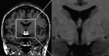
Figure 1 Magnetic resonance image acquisition of the whole-head coronal slice. Segmentation of the hypothalamus is shown in white. |
(suppressing testosterone virtually totally) after a 4-month interval compared with male comparison subjects, who showed a small increase. In addition, these effects were due to a lack of change in hypothalamic volume in testosterone-treated FMs after a 4-month interval, whereas it decreased in female comparison subjects. Moreover, testosterone administration to FMs increased total brain volume, whereas it remained unchanged in female comparison subjects. |
Moreover, testosterone administration to FMs increased total brain volume, whereas it remained unchanged in female comparison subjects. Significant group!sex! time interactions were also found for third (F(1,23)Z 16.56, P!0.0001) and lateral (F(1,23)Z21.90, P! 0.0001) ventricle volumes. The effects were due to increases in ventricle volumes in MFs as compared with a lack of change in third ventricle volume and subtle increase in lateral ventricle volume in male comparison subjects. Moreover, the findings were due to decreases in third and lateral ventricle volumes in FMs as compared with no change or subtle increases in volume in female comparison subjects. No significant effects were found for overall cerebral gray and white matter volumes. |
In the young adult control subjects, no significant changes in volumes over time were found, except for an increase in lateral ventricle volume irrespective of sex (F(1,11)Z5.67, PZ0.036). Intracranial volume showed a small (and insignificant) increase in volume of 0.2%. Although not significant, hypothalamus volume in female controls decreased over time. |
As expected, at first measurement, intracranial volume was significantly larger in males as compared with females irrespective of the condition of transsexualism (F(1,24)Z10.10, PZ0.004). No differences in intracranial volume were found between transsexuals and comparisons. After controlling for intracranial volume at initial measurement, no significant differences between the sexes or between transsexuals and control subjects were found for any of the brain volume measures. The relative proportion of white matter to gray matter was somewhat larger in the |
control females and the relative proportion of gray matter was somewhat larger in the control males, whereas the opposite was found for the transsexuals. However, this finding did not reach statistical significance. Third ventricle volume was larger in the transsexuals than in the controls, although not significant (main effect for group: F(1,23)Z3.36, PZ0.08; Table 1, Fig. 3). Thus, it is unlikely that the sexual dimorphic effects of sex hormone treatment on brain volumes over time were due to initial differences between groups or sexes. Moreover, it is also unlikely that level of education or scan interval influenced the results. Adding these factors to the analyses did not alter the results. |
The findings suggest that treatment of MFs with estrogens and anti-androgens decreases the male brain size towards female proportions, whereas treatment of FMs with androgens (not substantially affecting circulating estrogen levels) increases the female brain size towards male proportions. The magnitude of this change (i.e. 31 ml over a 4-month period) is striking, since it signifies a decrease in brain volume, which is at least ten times the average decrease of around 2.5 ml per year in healthy adults (16). Moreover, please note that consistent with the findings in the young adult control subjects of the present study, the longitudinal brain volume changes in young adults are normally small as compared with the older adults (17). The changes in total brain and hypothalamus volumes following cross-sex hormone treatment in the transsexuals were mirrored by changes in their third and lateral ventricle volumes, i.e. treatment with estrogens and anti-androgens in MFs increased third and lateral ventricle volumes, whereas treatment with androgens decreased the third and lateral ventricle volumes in FMs. This suggests that the total brain volume changes are at least in part due to changes in medial brain |
|
|